|
Micropaleontology The Study of Microscopic Fossils
|

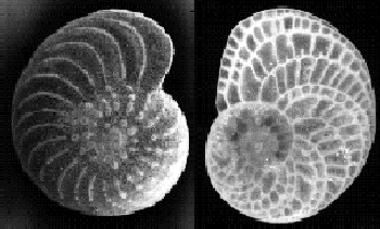
Foraminifera (foram): photo from Texas A & M University ODP
|
 Foraminifera (foram): photo from Texas A & M University ODP |
|
Microscopic animals and plants of the oceans can tell much about an area. Foraminifera (known as forams!), radiolaria (affectionately called rads!), nannoplankton, and diatoms were studied extensively aboard the Glomar Challenger. Forams can actually be seen with the naked eye, appearing to be a sand grain. They fizz when they dissolve in acid as they are made from calcareous material. They also can be crunched by your teeth (they are little shells) which can distinguish them from sand. Mrs. Paluso had the honor of having Dr. Hans Bolli name a foram after her when he discovered many new species aboard the Glomar Challenger. Radiolaria are smaller than forams. They are a group of unicellular organisms that are more often studied under a microscope than seen in real life. As living creatures they can be found in the great oceans but more commonly in the Central Pacific. They were discovered in strange and beautiful forms by the Glomar Challenger as a huge deposit of ooze on the ocean bed. Mrs. Paluso first starting preparing rads for Dr. Bill Riedel, at Scripps Institute of Oceanography, while she was an undergraduate at UCSD. Radiolaria may be seen with the naked eye as tiny clear bumps on a wet fine screen. But when dry multitudes of rads together can resemble spun glass fluff. The most striking feature of the Radiolaria is the beautiful glass like skeleton. Check out links for photos! In addition to forams and radiolaria, other fossils are also used to determine the age of sediments or for the study of the ocean floor. You may find PHOTOS and information to the radiolaia (Rads!), foraminifera (Forams!), Nannoplanton, Diatoms, and Ostacods by checking out the links. |
Links: |